Long COVID: Diagnosis of Post-acute Cardiovascular Sequelae of COVID-19
European Heart Journal:
Worldwide, survivors of COVID-19 now exceed hundreds of millions, with some reporting incomplete recovery months beyond the acute illness, a condition commonly referred to as long COVID. Persistent symptoms of breathlessness, chest pain, fatigue, headaches, brain fog, and palpitations are a constant reminder of the devastation caused by this virus and the need to remain vigilant for any long-term damage.
Proposed model for investigation of cardiovascular sequelae and long COVID
An approach considered reasonable by some experts involves screening of high-risk individuals for ongoing cardiac involvement including those with abnormal cardiac investigations during the acute phase, new CV diagnosis post-COVID-19, and athletes.
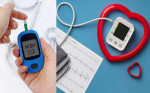
Screening of high-risk individuals could comprise of a thorough history, clinical examination, blood test panel (C-reactive protein, troponin, B-type natriuretic peptide/NT-proBNP, glycated haemoglobin, and lipids), ECG, and transthoracic echocardiography at least 8–12 weeks from infection.
For patients with clinically significant abnormalities after the screening, additional testing is recommended.
ECG: COVID-19 patients (e.g. with ongoing inflammation) may potentially benefit from ECG monitoring in the long term. Helpful for the diagnosis of atrial and ventricular arrhythmia, orthostatic tachycardia syndrome and ischaemic ECG changes during exercise.
Echocardiography: Echocardiography is vital in the early diagnosis of cardiac pathology in COVID-19 infection (suspected myocarditis, Takotsubo syndrome, myocardial infarction, pericardial effusion, etc.), particularly where haemodynamic stability is uncertain.
Magnetic Resonance Imaging (MRI): Diagnosis of myopericarditis, myocardial infarction, cardiomyopathy, monitoring of disease progression and recovery of tissue abnormalities, assessment of biventricular function, assessment of micro and macrovascular function, assessment of diastolic function.
Cardiopulmonary exercise test: Objective assessment of exercise capacity and contributing factors to delineate the predominant cause (cardiac, pulmonary, skeletal muscle, anemia) for exercise intolerance. Enables assessment of autonomic response during recovery.
CT pulmonary and coronary angiography: Computed tomography (CT) angiography (CTA) can detect pulmonary and coronary thrombosis, vasculitis, perivascular inflammation and thrombus burden in large vessels, epicardial coronary stenoses, or vascular pathology (e.g. mural thrombus or vasculitis) related to acute SARS-CoV-2 infection.
Cardiac SPECT: Provides function information/haemodynamic significance of coronary stenosis (micro and macrovascular).
Tilt Table test: Useful in the diagnosis of Postural orthostatic tachycardia syndrome (POTS), orthostatic hypotension, neurogenic syncope, vasovagal syncope.
CT computed tomography; ECG electrocardiography; POTS Postural Orthostatic Tachycardia Syndrome; SPECT Single Photon Emission Computed Tomography; CCTA coronary computed tomography angiography; CMR cardiac magnetic resonance; ESC The European Society of Cardiology; AHA The American Heart Association; EMB Endomyocardial Biopsy


Comments
You must login to write comment